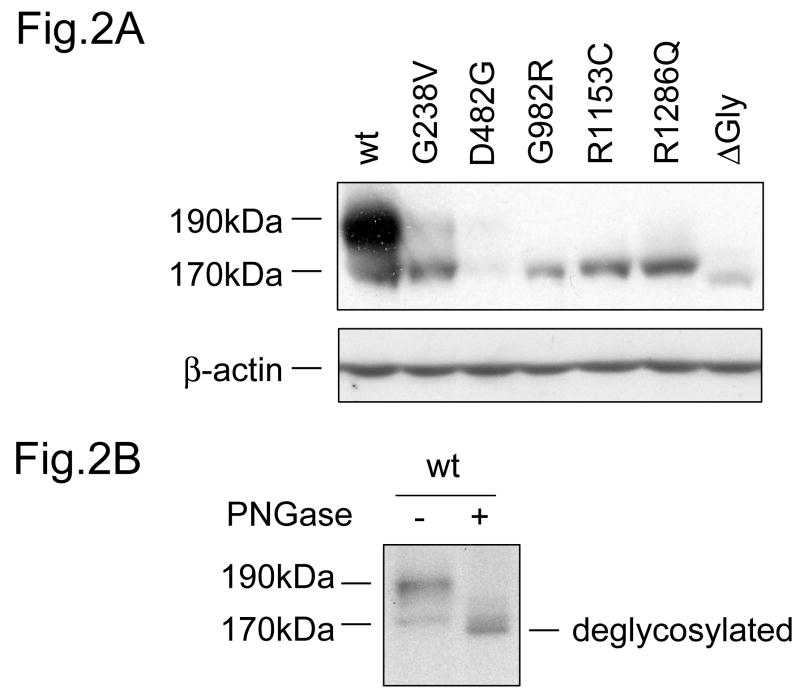
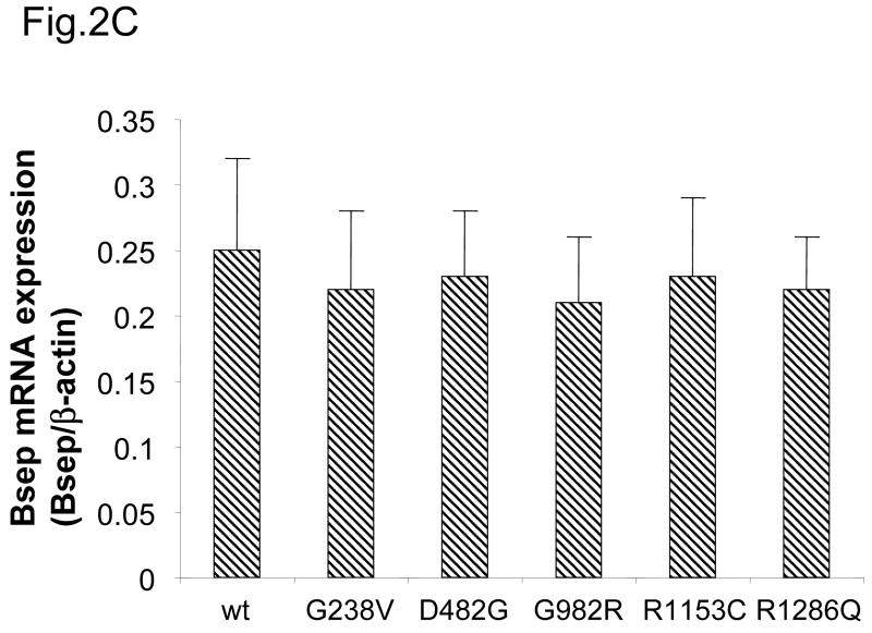
Fig.2. Expression of PFIC II mutants and ΔGly in HEK 293 cells.
(A) The HEK 293 cells were transiently transfected with wt GFP-Bsep, PFIC II mutants and ΔGly. The lysates (100 μg protein each lane) were separated by SDS-PAGE and GFP-Bsep was detected by immunoblotting using an antibody against GFP. β-actin was also probed to indicate the equal loading of lysates. ECL was used to visualize the protein of interest. 190 kDa and 170 kDa indicate the mature and immature glycosylated GFP-Bsep. (B) The cell lysate of HEK cells transfected with wt GFP-Bsep was digested with PNGase F. (C) mRNAs were isolated from the HEK cells 24 hrs after transfection. Real-time quantitative PCR was performed and data represent triplicate measurements for each sample. Bsep gene expression was normalized to the expression of β-actin.


