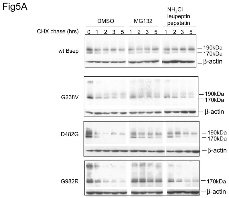
Fig.5. Proteasome is the major pathway to degrade PFIC II mutants.
(A) The HEK 293 cells were transfected with the wt GFP-Bsep, G238V, D482G, and G982R. Cycloheximide was added to the cell culture 24 hrs after transfection and the cells were harvested at indicated time points. GFP-Bsep in each cell lysate was analyzed by immunoblotting using an antibody against GFP. β-actin was also probed to indicate the equal loading of lysates. To inhibit proteasome, 20μg/ml MG132 was added to the culture medium during the cycloheximide chase. Equal amount of DMSO, a solvent used to dissolve MG132, was added to the control medium. To inhibit lysosome, 15mM ammonium chloride, 10μg/ml leupeptin and 10μg/ml pepstatin was added to the medium. ECL was used to visualize the protein of interest. The HEK 293 cells were transfected with wt FLAG-Bsep (B) and G982R (C). The cells were incubated with medium with DMSO (mock) or with 20μg/ml MG132 during the chase period. FLAG-Bsep was immunoprecipitated. Each data point represents mean ± SE of triplicate determination.


