
Figure 1.
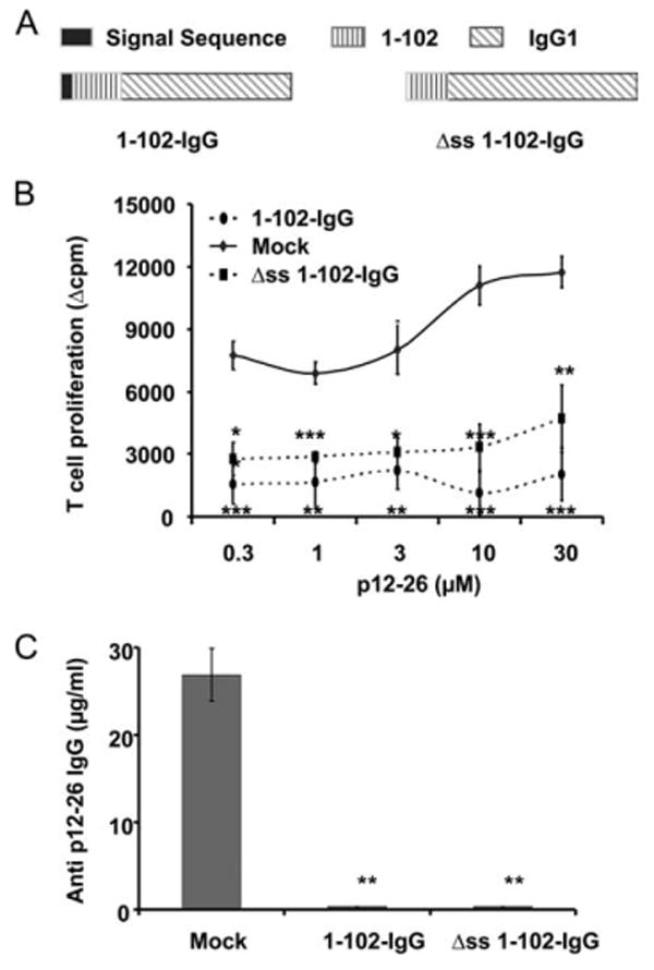
Secretion of peptide-IgG is not required for tolerance induction. A, The 1–102 cDNA was subcloned into the BSSK-IgG1 cassette, and then the 1–102-IgG1 fusion gene was subcloned into the MBAE retroviral vector. The signal sequence of IgG H chain was deleted to generate the Δss 1–102-IgG construct. B, Normal BALB/c recipients were injected i.p. with 107 Δss 1–102-IgG B cells, 1–102-IgG B cells, or an equal number of OVA-IgG B cells (mock control). On day 7 postinjection, animals were immunized in a hind footpad, and the base of tail with 25 μg of 12–26 peptide emulsified in CFA. Two weeks later, animals were sacrificed and T cells were isolated from draining LNs and assayed by [3H]thymidine incorporation. Data represent mean Δcpm ± SE for three or four animals. The background [3H]thymidine incorporation was in the range of 5,000–10,000 counts. This is representative of three independent experiments. ***, p < 0.001; **, p < 0.01; *, p ≤ 0.05. C, Animals were treated as described in B. Sera were collected 2 wk after immunization and analyzed by ELISA for total anti-12–26 IgG. B3.11 Ab was used as standard. This is representative of three independent experiments. **, p < 0.01.