
Figure 3.
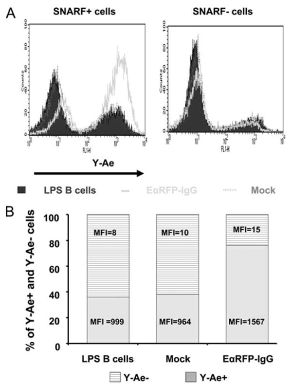
The Eα52–68:I-Ab complexes are derived from endogenous, but not exogenous, EαRFP-IgG. A, C57BL/6 B cells were stimulated with LPS for 24 h and then transduced with either EαRFP-IgG or HEL-IgG as a mock control. After 24-h coculture with irradiated packaging cells, B cells were collected and centrifuged over Lympholyte M to remove dead cells and then labeled with an intracellular dye SNARF (5 μM). SNARF-labeled untransduced LPS blasts, HEL-IgG, or EαRFP-IgG B cells were mixed with unlabeled, untransduced LPS B cells at 1:1 ratio. A total of 107 mixed cells was cultured in 5 ml of medium in 6-well plate. After 24 h, cells were collected and stained with biotin-Y-Ae mAb, followed by FITC-streptavidin. SNARF+ and SNARF− cells were gated and presented. Non-transduced LPS B cells are depicted by the solid histogram; HEL-IgG-transduced B cells by the dotted line; EαRFP-IgG-transduced B cells by the thick line. B, The percentages and mean fluorescence intensity (MFI) of Y-Ae+ and Y-Ae− cells in SNARF+ cells are indicated.