
Figure 3. Colocalization of internalized fibronectin with α5 integrins.
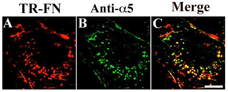
SMCs were incubated with 10 μg/mL TR-fibronectin and 50 μM chloroquine for 8 hours. Cells were stained using an anti-α5 integrin antibody (AB1928). A, TR-fibronectin; B, α5 integrin; C, overlay image. These images are optical sections collected from a confocal microscope. Scale bar, 10μm.