
Figure 7. Reduced endocytosis and turnover of pre-assembled fibronectin matrix in β1-integrin-null cells.
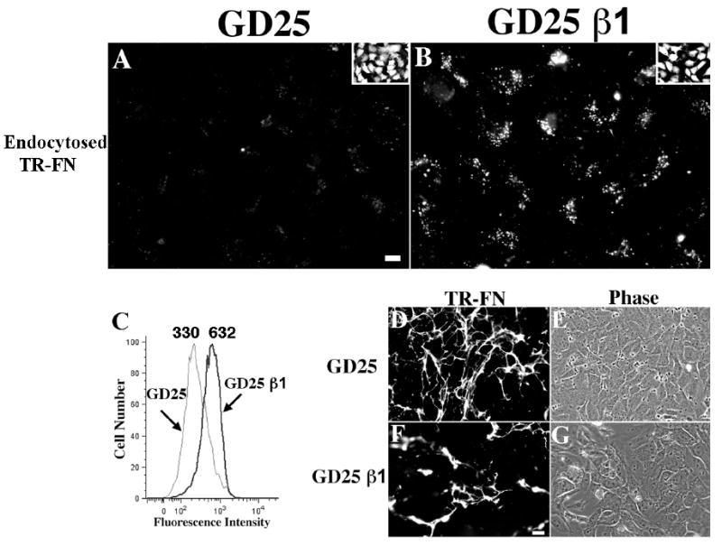
GD25 and GD25 β1 re-expressing cells were seeded on pre-assembled TR- (A, B, D, E, F, G) or AF488 (C)-fibronectin matrix and incubated for 24 hours at 37°C. (A-B) Cells were incubated with 0.2% trypan blue for 3 minutes and then fixed. Intracellular fibronectin vesicles are shown (A, GD25; B, GD25 β1). The insets show outlines of cells loaded with CellTracker Green. Scale bar, 10μM. (C) Cells were processed for flow cytometry to quantitate endocytosed fibronectin. The numbers over the peaks in C are the MFI of internalized AF 488-fibronectin. (D-G) Cells were fixed without trypan blue treatment to visualize fibronectin fibrils (D, E, GD25; F, G, GD25 β1; E and G are phase images of corresponding fields). Scale bar, 40μm.