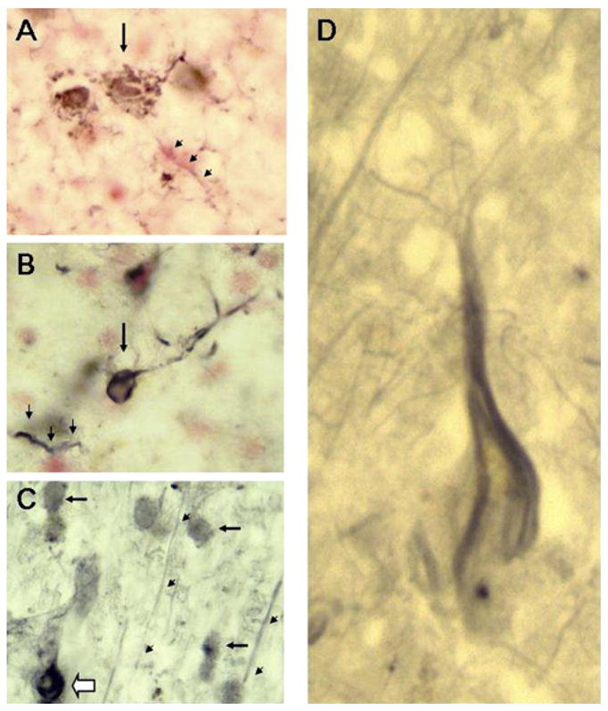
Figure 3. Gab2 Colocalizes with Dystrophic Neurons in the AD Brain.

(A) LOAD hippocampus (neutral red counterstain) (40× objective). The arrow indicates a highly dystrophic cell with the size and morphology of a cortical pyramidal neuron. Arrowheads point to one of many structures in the sections that resemble dystrophic neurites or neuropil threads. (B) LOAD hippocampus (neutral red counterstain) (40×). The arrow denotes a putative neurofibrillary tangle-containing neuron. Arrowheads again indicate a dystrophic neurite. (C) LOAD posterior cingulate gyrus (40×). Filled arrows point to normal-appearing putative neurons. The open arrow points to a cell with the features of a neurofibrillary tangle-bearing neuron. Immunoreactive structures clearly resembling pyramidal cell apical dendrites were also observed ascending through the cortical layers (arrowheads). (D) LOAD posterior cingulate cortex (100× objective). Shown is a Gab2 immunoreactive cell with the flame-shaped cytoplasmic inclusion typical of the neurofibrillary tangle.
