
Figure 6. Adp Heterozygous Mutant Mice Are Obese and Insulin Resistant.
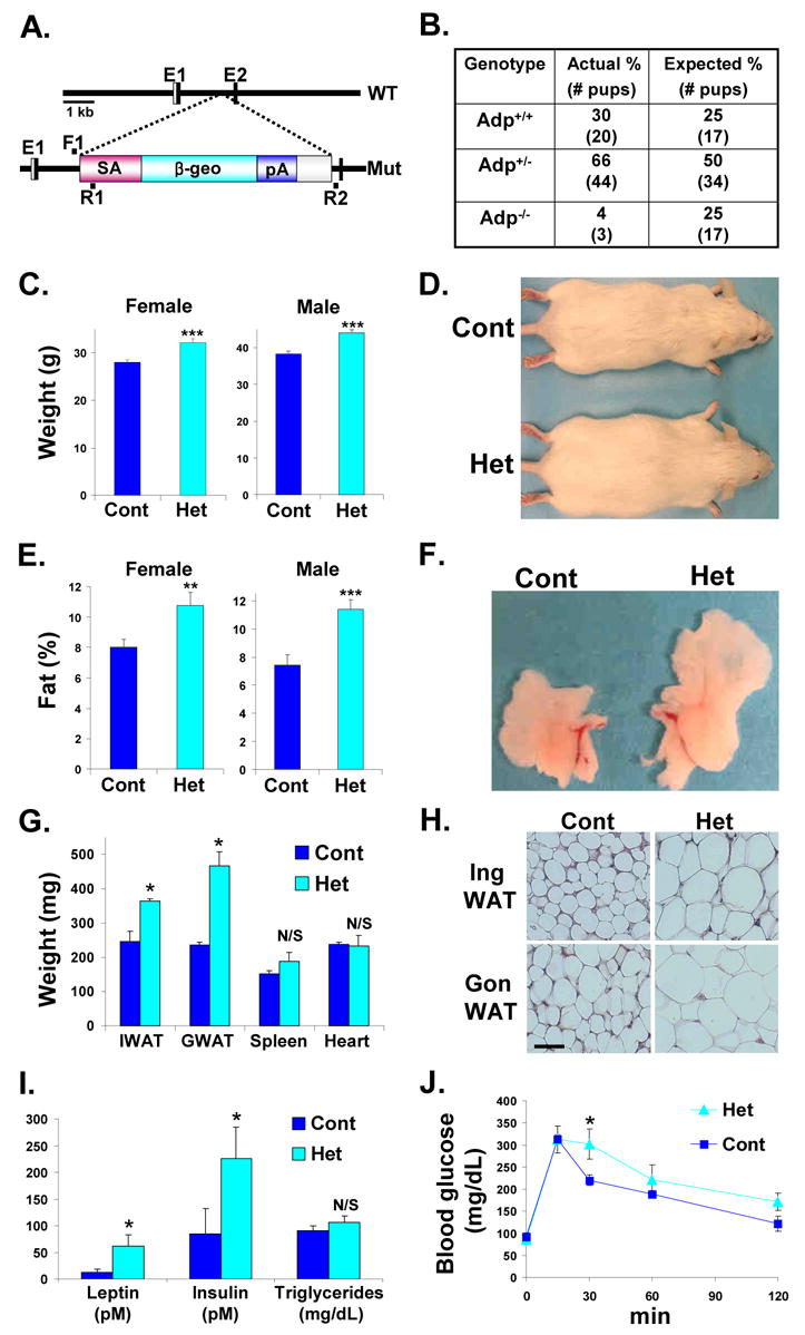
(A) Schema of the wild-type (WT) Adp locus and the mutant (Mut) Adp allele. Splice Acceptor (SA) and the lacZ-neomycin (β-geo) cassette were inserted and disrupt gene expression from the second exon (E2). The location of the genotyping primers (F1, R1, R2) are illustrated.
(B) Genomic DNA was extracted from pups of multiple Adp+/− by Adp+/− intercrosses and genotyped for the presence of Adp mutant and wild-type alleles.
(C) Average weights of littermate matched female (control n = 10 mice, Adp Het n = 12) and male (control n = 15, Adp, Het n = 20) cohorts.
(D) Photograph of representative wild-type (Cont) and Adp+/− mice (Het).
(E) Average fat content as assessed by NMR of littermate matched female (control n = 10 mice, Adp Het n = 12) and male (control n = 15, Adp Het n = 20) cohorts.
(F) Photograph of representative perigonadal white adipose tissue (WAT) explants from sibling wild-type (Cont) and Adp+/− (Het).
(G) Average weights of inguinal (I) and perigonadal (G) WAT and indicated organs of wild-type and Adp heterozygous littermates.
(H) Histological analyses of inguinal (Ing) and perigonadal (Gon) WAT.
(I) Plasma of control and Adp heterozygous cohorts was analyzed for leptin, insulin and triglyceride levels.
(J) GTTs of Adp heterozygous and control siblings (n = 8). Analyses in panels C–J done on 4-month old mice.
*p < 0.05, **p < 0.01, ***p < 0.005 by t-test. N/S not significant. Error bars indicate SEM.