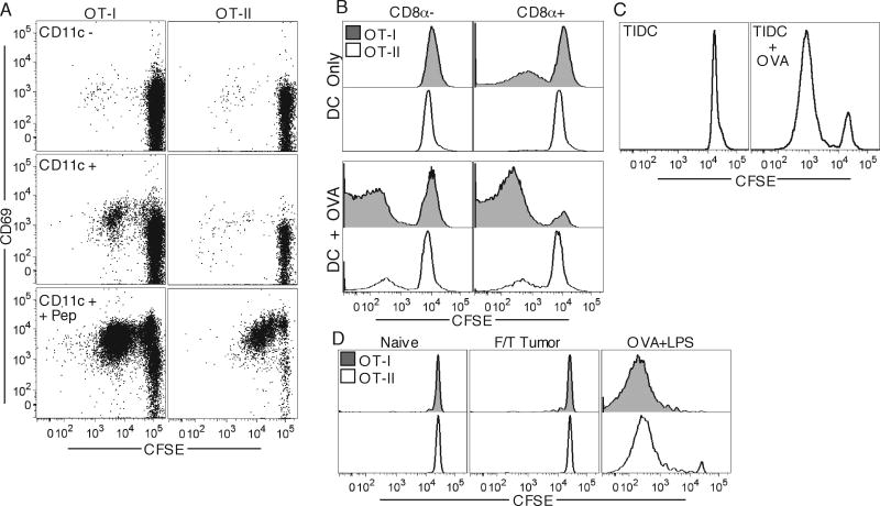
Figure 7. MHC-I but not MHC-II presentation of TAg by DC.
OT-I and OT-II T cells were in vitro incubated with CD11c– (top) and CD11c+ (middle, bottom) purified cell fractions from DLN of day 12 B16.OVA-bearing animals having OVA257-264, 323-339 peptides added to some wells (bottom), and were analyzed for CFSE dilution and CD69 expression four days later (A). OT-I and OT-II T cells were incubated with CD8- (left) and CD8+ (right) purified DC fractions from DLN of day 13 B16.OVA-bearing animals with OVA protein added to some wells (bottom), and were analyzed for CFSE dilution four days later (B). OT-II T cells were incubated with tumor-derived purified CD11c+ cells (TIDC) with (right) or without (left) addition of OVA protein (C). OT-I and OT-II T cells were adoptively transferred into recipients which were challenged a day later with 107 freeze/thaw killed tumor cells or 100μg OVA protein and LPS. DLN were harvested four days later and analyzed for T cell CFSE dilution (D).

