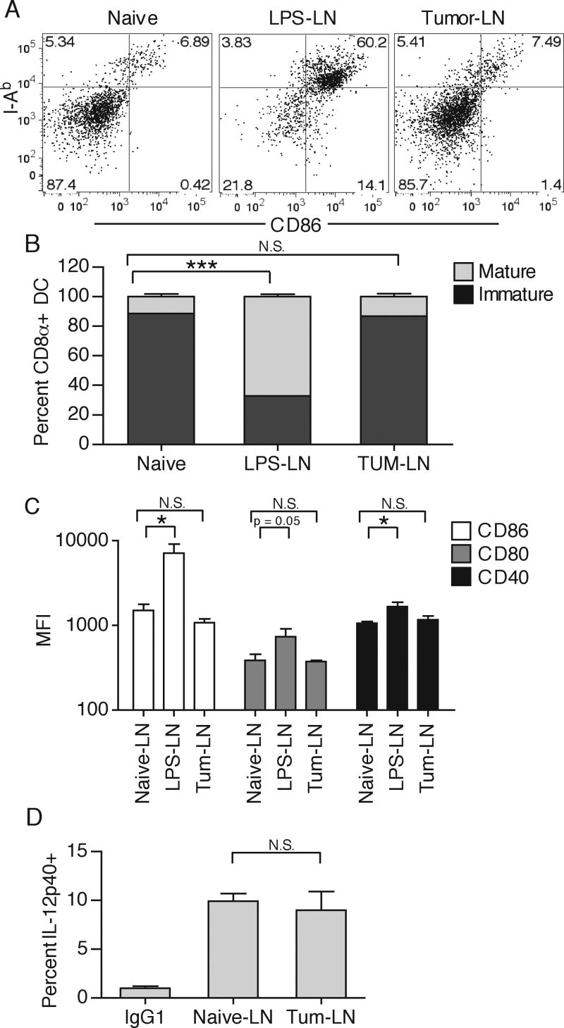
Figure 8.
Inguinal DLN from either naïve, LPS-treated (50μg subcutaneously injected 16 hours prior), or day 12 B16.F10-bearing mice were collected and CD8α+ DC were examined for expression of I-Ab and CD86 by gating on CD3-, CD19-, CD11c+, CD11b-, CD8α+ cells (A). Immature and mature CD8α+ DC were quantified by counting percent I-Ab positive/dim or I-Ab positive/bright, respectively (B). CD8α+ DC mean fluorescence intensity (MFI) for CD86, CD80, and CD40 was quantified (C). Percent IL-12p40+ of total CD8α+ DC was quantified after a 4hr Brefeldin A in vitro incubation. No enhancement in IL-12p40 production was detected in the LPS-treated group. (D). Results are represented as mean ± SD. Data is representative of at least two independent experiments.

