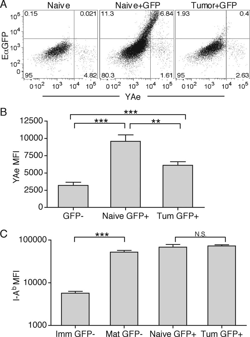
Figure 9. Inhibition of migratory DC MHC-II presentation in DLN.
Naïve or day 10 B16.F10 tumor-bearing animals were s.c. or i.t. (respectively) injected with 50μg EαGFP. 24 hours later, CD11c+ DC were isolated from DLN and separated into resident and migratory DC subsets. Migratory DC (gate: CD3-, CD19-, I-Ab+, CD11c+, CD11b+, CD8α-, CD4-) were then analyzed for EαGFP fluorescence and YAe staining (A). GFP- and GFP+ migratory DC from naïve or tumor-bearing (Tum) DLN were analyzed for total YAe MFI, mean fluorescence intensity (B). Numbers represent percent of total cells in each quadrant. GFP- CD11b+ DC were separated into immature I-Ab positive/dim (GFP- Imm) and mature I-Ab positive/bright (GFP-Mat) subsets and compared to GFP+ migratory DC from naïve or tumor-bearing DLN for MFI of I-Ab (C). Results are represented as mean ± SD. Data are representative of two independent experiments

