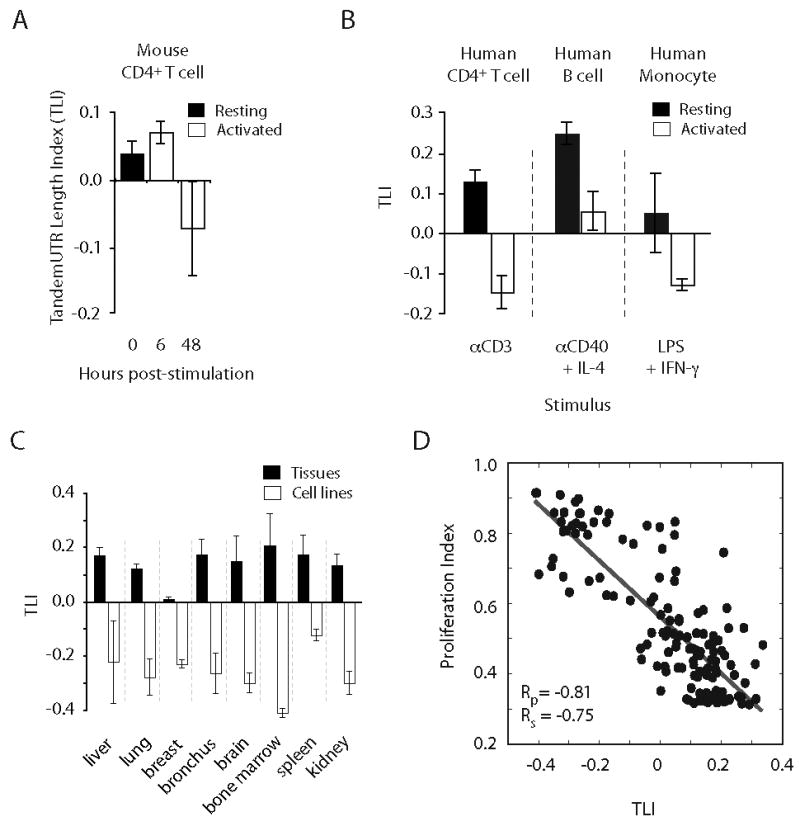
Figure 2. Reduced expression of extended 3′ UTR regions is widespread, conserved to human, and correlated with proliferation.

(A) TLI analysis of murine CD4+ T lymphocytes stimulated with anti-CD3/anti-28 beads (this study). (B) TLI values from human CD4+ T cells, B cells, and monocytes activated for 30 h with the indicated stimulus. (C) TLI values of cultured cancer cell lines and of matched normal tissues. (D) Correlation of TLI with proliferation index across a panel of 135 different tissues and samples (Rs = Pearson; Rp = Spearman correlations). Data in (A-D) are mean +/- SD of 2-33 replicates (Table S8).
