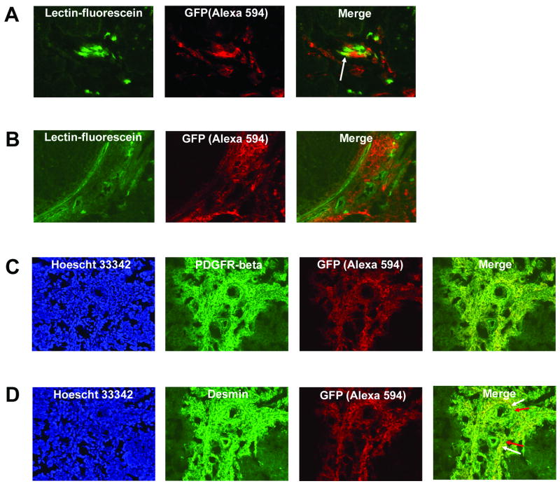
Fig. 1.
Bone marrow cells differentiate into both endothelial and vascular smooth muscle cells within perfused, functional Ewing’s tumor neovessels. (A) TC71 Ewing’s tumors were implanted in mice previously transplanted with GFP+ BM. Donor-derived cells were detected by staining tumor sections with anti-GFP, followed by a secondary antibody conjugated to the Alexa 594 fluorophore (red). Functional tumor vessel endothelium was labeled green by intravascular perfusion with fluorescein-conjugated lectin. The arrow in the merged image indicates BM cells that differentiated into endothelial cells within perfused tumor vessels (yellow). Images were analyzed using a 20× objective. (B) Migrated BM cells (red) reside in close association with numerous lectin-marked functional TC71 tumor neovessels (green). (C, D) TC71 tumor sections were co-stained for GFP+ BM cells and either PDGFRβ (C) or desmin (D). Extensive networks of PDGFRβ+ (1C) and desmin+ (1D) cells are seen throughout the tumor area. The merged images indicate that BM-derived cells express PDGFRβ and desmin (denoted by yellow fluorescence), suggesting vascular smooth muscle cell differentiation. Hoescht 33342 staining confirmed that marker expression was associated with cells. The tumor areas shown were analyzed with a 20× objective. Arrows (Fig. 1D) indicate that the vascular smooth muscle cell network is a mosaic of locally-derived and BM-derived cells.

