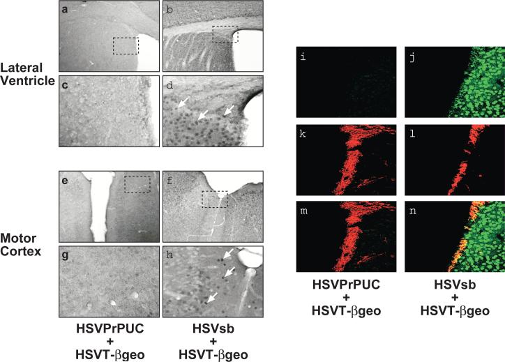
Figure 1. In utero co-delivery of HSVT-βgeo and HSVsb to E14.5 mouse CNS results in extensive transgenon expression throughout the mouse brain.
At 29 days of age, inoculated animals were sacrificed, perfused with 4% paraformaldehyde, brain sections processed for LacZ/Diaminobenzidine (DAB) immunohistochemistry, and sections imaged using light microscopy (n=4 per treatment group). Representative photomicrographs of the lateral ventricle (a-d) and motor cortex (e-h) from mice receiving HSVPrPUC + HSVT-βgeo (a, c, e, and g) and HSVsb + HSVT-βgeo (b, d, f, and h) are depicted. Magnification = 10X in panels a, b, e, and f. Hatched boxes shown in a, b, e, and f represent regions magnified in c, d, g, and h, respectively (final magnification = 40X). White arrows highlight LacZ-immunopositive cells. Adjacent coronal brain sections were processed for dual LacZ/DCX fluorescent immunohistochemistry and imaged using confocal microscopy. Expression of LacZ in the mice was detected in the green channel (i, j). Neuronal progenitor marker DCX, expressed in migrating neuronal progenitors lining the sub-ventricular zone and associated with the rostal migratory pathway in adult mice, was detected in the red channel (k, l). Co-localization of LacZ with the DCX signal is depicted in yellow (m, n).

