
Figure 1. αv integrin dimerized with β3 integrin in rat astrocytes.
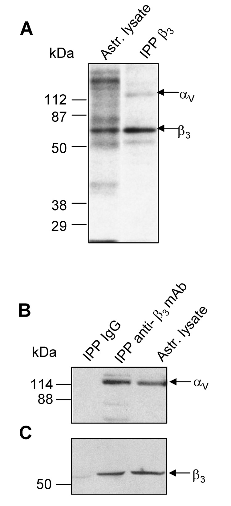
A) Surface biotinylated proteins were immunoprecipitated (IPP) with anti-β3 integrin mAb. Labelled proteins from astrocyte lysates and IPP proteins were revealed with streptavidin-HRP. B) and C) Proteins in a whole cell lysate were precipitated with anti-β3 mAb or a control IgG. IPP complexes were separated by SDS-PAGE in parallel with astrocyte lysate. Proteins transferred to nitrocellulose were revealed with anti-αv mAb (B) and anti-β3 pAb (C). Values for molecular mass in kDa are shown to the left of each panel.