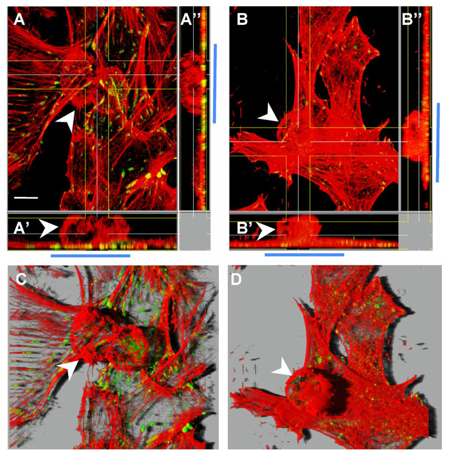
Figure 4. The formation of focal adhesions but not neuron-astrocyte interaction per se was completely dependent on Thy-1-integrin binding.

Astrocytes were washed with serum-free medium and stimulated with CAD cells either not treated (A and C) or treated with hybridoma supernatant containing anti-Thy-1 mAb (clone V8) (B and D) as indicated in Figure 3. After 10min of neuron-astrocyte incubation, cells were gently washed and then prepared for immunofluorescence analysis of focal adhesions (green) and F-actin (red). White arrowheads indicate CAD cells bound to a monolayer of astrocytes. These are easier to observe in orthogonal sections that show cells in the xz plane (A’ and B’) and xy plane (A’’ and B’’). Blue bars mark the area where CAD cells (round cells) were bound to the astrocyte monolayer. In A’ and A’’ green/yellow dots corresponding to focal adhesions are apparent in the area of cell-cell contact. In B’ and B’’, on the other hand, very few and small focal points are detectable in the area of contact. C and D correspond to the same fields shown in A and B, respectively. However here, z-stack-sections are visualized in 3D projections obtained using Imaris software (Bitplane AG, Zurich, Switzerland) to show that CAD cells bound to the astrocyte monolayer induced focal adhesion and stress fiber formation only when Thy-1 was available (C) and not in the presence of an anti-Thy-1 antibody (D). Results shown are representative of 3 individual experiments performed in duplicates. Magnification bar is 10µm.
