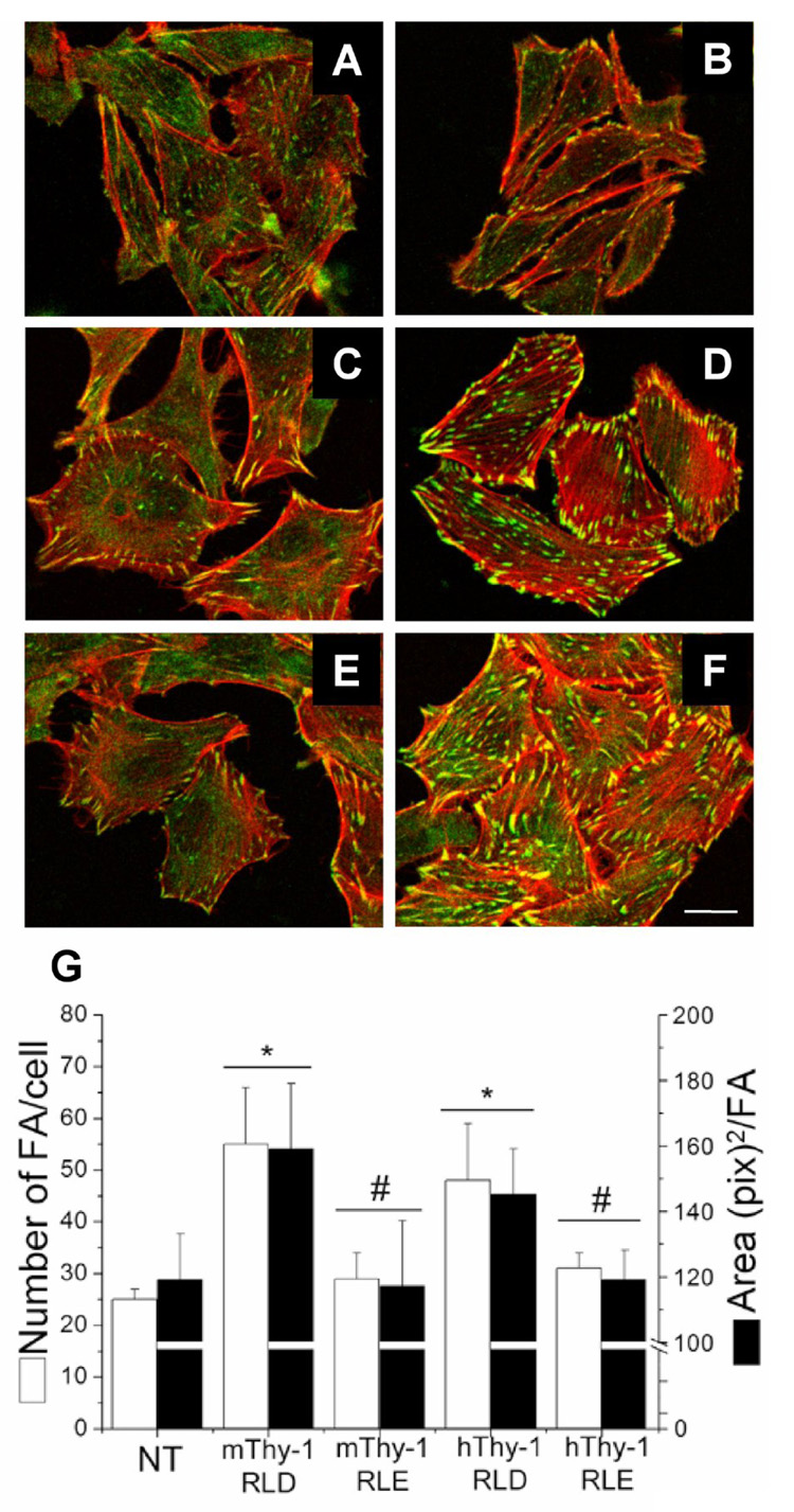
Figure 7. Mouse and human Thy-1-Fc wild type proteins stimulated focal adhesion and stress fiber formation in rat astrocytes.

Astrocytes either not treated (A) or treated for 10min with Protein-A Sepharose beads containing TRAIL-R2-Fc (B), mouse Thy-1(RLE)-Fc (C), mouse Thy-1(RLD)-Fc (D), human Thy-1(RLE)-Fc (E) or human Thy-1(RLD)-Fc (F) were fixed, permeabilized and stained for focal adhesions with anti-paxillin mAb (green) and stress fibers with Rhodamine-coupled phalloidin (red). G) Number of focal adhesions/cell (white bars), as well as the average area/focal adhesion (black bars) were quantified using Image J software from NIH public domain. Only mouse and human Thy-1(RLD)-Fc significantly stimulated the formation of new and larger focal adhesions. Images are representative of results obtained in at least 3 different experiments. *, p < 0.05 with respect to the control non-treated (NT) cells. #, p < 0.05 compared to the values obtained when stimulating with the respective mouse or human RLD protein. Magnification bar is 20µm.
