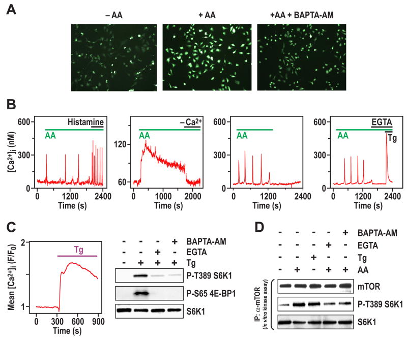
Figure 2. AA stimulation evokes an increase in [Ca2+]i. (A).
Confocal images of Fluo-4 fluorescence stained HeLa cells treated with AAs as in Figure 1A for 5 min in the absence or presence of BAPTA-AM. (B) [Ca2+]i traces in fura2-loaded HeLa cells stimulated with AAs as in Figure 1A. First panel shows representative single-cell [Ca2+]i response to AAs followed by 0.5 μM histamine addition. Second panel shows effect of removing extracellular Ca2+ by replacement of medium with AA-containing buffer without added Ca2+. Third panel shows effect of AA withdrawal on single-cell [Ca2+]i response; this experiment was carried out in the presence of 10% dialyzed FCS. Fourth panel shows effect of extracellular Ca2+ chelation with 4 mM EGTA, followed by treatment with 2 μM Tg.. (C) Left panel, HeLa cells plated onto 35 mm tissue culture plates were deprived of serum and AAs, then loaded with Calcium Green-1/AM and [Ca2+]i responses were monitored by confocal microscopy before and after the addition of 150 μM Tg. The trace is the mean Tg-evoked [Ca2+]i increase (n = 332 cells). Right panel, HeLa cells treated with AAs as in Figure 1A, then with 150 μM Tg for 30 min in the absence or presence of either 2 mM EGTA or 2 μM BAPTA-AM. EGTA and BAPTA-AM were added 15 min prior to the addition of Tg. (D) HeLa cells treated with either EGTA or BAPTA-AM were stimulated with AAs or Tg for 15 min as described in Figures 1A and 2C, respectively. Cells were harvested, lysed, mTOR was immunoprecipitated and in vitro mTORC1 activity was assayed as described (Sancak et al., 2007).

