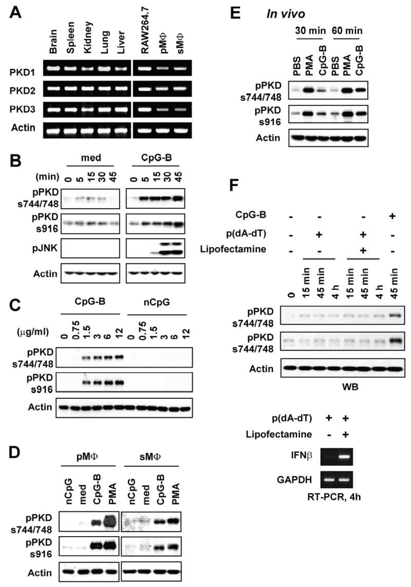
Figure 1. CpG-B DNA induces phosphorylation of PKD family members in macrophages.

A, Levels of the indicated mRNA in various tissues, RAW264.7 cells and primary macrophages isolated from C57BL/6 mice were detected by RT-PCR analysis. PMΦ: peritoneal macrophages, sMΦ: splenic macrophages. B–D, RAW264.7 cells (Panels b and c), peritoneal (pMΦ) or splenic (sMΦ) macrophages isolated from C57BL/6 mice (Panel d) were stimulated with medium (med), CpG-B DNA (CpG-B), non-CpG DNA (nCpG), or PMA. Concentrations of DNA used are either indicated or 12 μg/ml. PMA was used at 10 ng/ml. Periods of DNA stimulation are either indicated or 45 min. Activation status of PKDs was detected by phospho-specific Western blot assay using Abs specific for the phosphorylated forms of PKD (pPKDs744/748, pPKDs916). E, BALB/c mice were injected intraperitoneally with PBS, CpG-B DNA (30 μg/mouse), or PMA (30 ng/mouse). At the indicated times later, peritoneal cells were isolated. Activation status of PKDs was detected by phospho-specific Western blot assay. F, RAW264.7 cells were stimulated with 6 μg of poly(dA-dT) in the presence or absence of lipofectamine or CpG-B DNA (6 μg) up to 4 h. Activation status of PKDs was detected by phospho-specific Western blot assay. Expression of IFN-β as a stimulation indicator was detected by RT-PCR.
