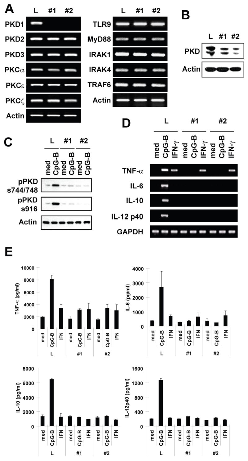
Figure 5. PKD1 plays an indispensable role in cytokine production in macrophages in response to CpG-B DNA.

A–C, RAW264.7 cells were stably transfected with vectors expressing control luciferase-shRNA (L) or PKD1shRNA (clone #1 and #2) under control of the H1 promoter. Positive transfectants were selected and maintained in selection medium containing G418 (1 mg/ml). Messenger RNA levels of selected genes in PKD family members, PKC family members, and TLR9 and its down-stream signaling modulators were analyzed by RT-PCR (A). PKD protein levels in gene-specific knockdown RAW264.7 cells were examined using Western blot analysis (B). Phosphorylation of PKD in gene-specific knockdown RAW264.7 cells after CpG-B DNA stimulation (12 μg/ml for 45 min) was detected by Western blot (C). D and E, Control luciferase-knockdown (L) or PKD1-knockdown (#1 and #2) RAW264.7 cells were stimulated with medium, CpG-B DNA (6 μg/ml) or IFN-γ (10 ng/ml) for 4 h (D) or 24 h (E). Levels of cytokine mRNA (D) and protein (E) were analyzed by RT-PCR and cytokine-specific ELISA, respectively. ELISA data represent the mean cytokine concentration (pg/ml) ± SD of triplicates.
