
Figure 8. CpG-B DNA induces activation of PKD1 through an endosomal pH-sensitive TLR9/MyD88/IRAK1-dependent signaling pathway.
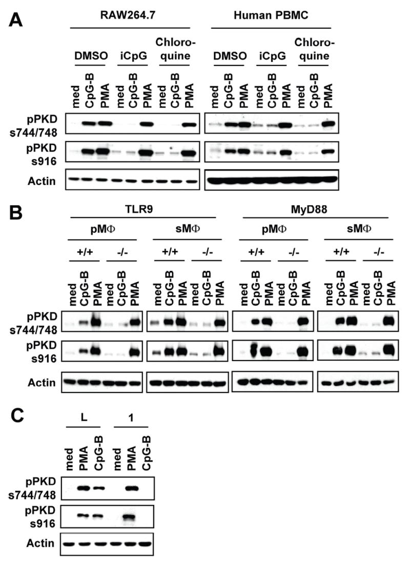
A, RAW264.7 cells and human PBMCs were pretreated with chloroquine (2.5 μg/ml) or iCpG DNA (12 μg/ml) for 30 min and then stimulated with medium (med), CpG-B DNA (12 μg/ml) or PMA (10 ng/ml) for 45 min. B, Peritoneal (pMΦ) or splenic (sMΦ) macrophages isolated from TLR9+/+ (BALB/c), TLR9−/−, MyD88+/+ (wild-type littermates), or MyD88−/− were stimulated with medium, CpG-B DNA (12 μg/ml) or PMA (10 ng/ml) for 45 min. Activation status of PKD1 was detected by phospho-specific Western blot assay. C, Control luciferase-knockdown (L) or IRAK1-knockdown (1) RAW264.7 cells were stimulated with medium, PMA, or CpG-B DNA for 45 min. Phosphorylation status of PKD1 was detected by Western blot analysis. Lack of IRAK1 expression in IRAK1-knockdown macrophages was confirmed (36).