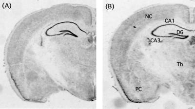
Figure 2.
Histological analysis. Histological sections containing hippocampus from Syt IV mutant (A) and wild-type (B) mice after staining with 0.5% thionin. Indicated are dentate gyrus (DG), pyramidal cells of the Schaffer collateral fiber tract (CA1 and CA3), thalamus (Th), neocortex (NC), and piriform cortex (PC).

