
Fig. 2.
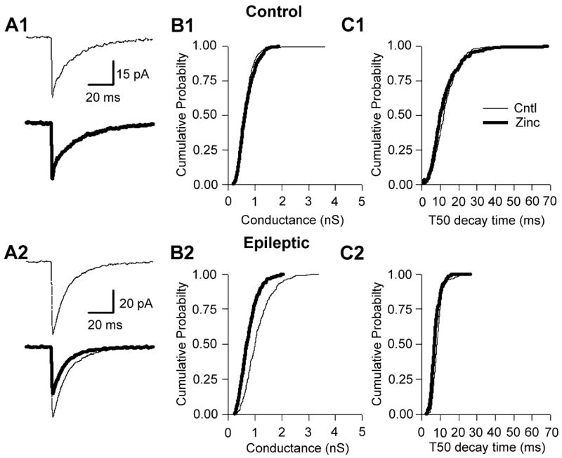
Zinc blocked mIPSCs in epileptic, but not control, DGCs. (A1) Averaged mIPSCs (50 events) taken before (thin line) and during (thick line) bath application of zinc (300 μM) to a control DGC. (B1) Cumulative probability–conductance and (C1) cumulative probability −50% decay time (T50) plots for the same cell as represented in A1, demonstrating that zinc has no effect on mIPSC conductance or T50 in control DGCs. (A2) Averaged mIPSCs taken before (thin line) and during (thick line) bath application of zinc to epileptic DGCs, demonstrating that zinc significantly reduced mIPSC amplitude (see A2 lower sweep; note difference in scale bar amplitude). (B2) Cumulative probability–conductance plot demonstrating a significant reduction in mIPSC conductance. (C2) However, the cumulative probability–decay plot for the same neuron as in A2 clearly demonstrates that zinc was without effect on epileptic mIPSC decay time.