
Fig. 6.
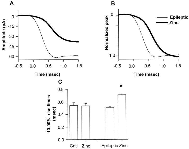
mIPSC kinetics were significantly altered during zinc exposure in epileptic neurons. (A) Averaged rise time (n = 20) from an epileptic DGC (thin line) in control aCSF (mean 0.47 ms), and in the presence of zinc (thick line, mean 0.76 ms). (B) Normalization of the zinc and control traces more clearly illustrates the slowed onset of mIPSCs during zinc exposure. The traces were lined up by aligning their 10% rise times. (C) Histogram of mean mIPSC 10–90% rise times in control and epileptic populations in normal and zinc-containing aCSF. *P ≤ 0.05, paired t-test, between epileptic in control and zinc-containing aCSF.