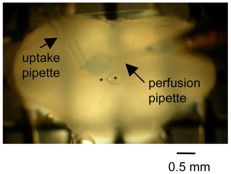
Figure 1.

Image of local unilateral perfusion. In this example, ACSF containing fast green was locally and unilaterally perfused onto the XII nucleus of a rhythmically active medullary slice preparation. Perfusion region is circumscribed to a zone approximately 0.5 mm in diameter as revealed by fast green labeled region over the left XII nucleus. Asterisks are over epoxy beads on a nylon thread that help to limit the labeled perfusion region. Arrows indicate the local perfusion pipette and the local uptake pipette.
