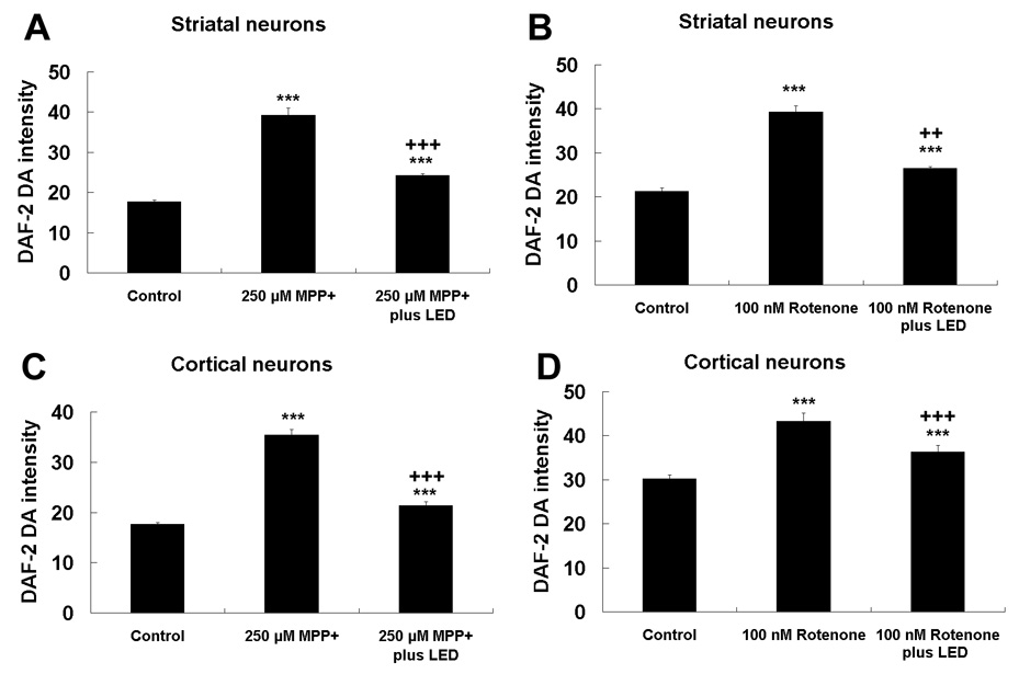
Fig. 9.

NO production measured by DAF-2 DA fluorescence in neurons exposed to MPP+ or rotenone, with or without twice-a-day LED treatment. A and C: Striatal and cortical neurons, respectively, exposed to MPP+; B and D: Striatal and cortical neurons, respectively, exposed to rotenone. DAF-2DA expression was increased by MPP+ or rotenone exposure but decreased by LED treatment. However, the fluorescent signal was still higher than control levels. ***, P < 0.001 compared to controls. All +P values were compared to MPP+ or rotenone only: ++, P < 0.01, +++, P < 0.001.
