
Figure 1. Mdm2 physically interacts with L26 in mammalian cells.
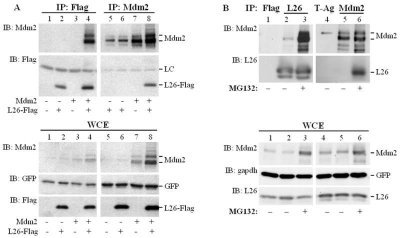
A. HEK293 cells (1.75×10⁶ cells/10cm dish) were transiently co-transfected with the indicated combinations of expression plasmids encoding L26-Flag (6μg/dish), wild type human Mdm2 (6μg/dish), and GFP (0.3μg/dish) as a transfection control. Cells were harvested and extracted 48 hours later. Top panel: Extracts were immunoprecipitated (IP) with anti-Flag or anti-Mdm2 antibodies, and the immunoprecipitated material was resolved by SDS-PAGE and subjected to Western immunoblot analysis (IB) with the indicated antibodies. LC: antibody light chain. Bottom panel: 2.5% of each whole cell extract (WCE) was resolved by SDS-PAGE and subjected to Western blot analysis with the indicated antibodies.
B. SJSA-1 cell cultures were treated with 8μM MG132 overnight where indicated, or left untreated. The next day, cells were harvested and subjected to protein extractions. Extracts were immunoprecipitated (IP) with control (anti-Flag rabbit polyclonal, upper panel, lane 1) or anti-L26 (lanes 2, 3) antibody (two 15cm dishes/lane). For the reciprocal reaction, extracts were immunoprecipitated with control (anti-SV40 large T antigen, lane 4) or anti-Mdm2 (lanes 5, 6) antibodies (four 15cm dishes/lane). The immunoprecipitated material was resolved by SDS-PAGE and subjected to Western blot analysis with the indicated antibodies (IB). Lower panel: 1.7% of each whole cell extract was analyzed directly by SDS-PAGE, followed by Western blot analysis (IB) with the indicated antibodies.