
Figure 3. Mdm2 promotes the proteasomal degradation of polyubiquitylated L26.
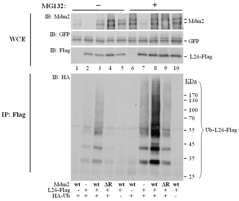
U2OS cells were seeded at 8×10⁵ cells/10cm dish. Twenty four hours later, cultures were transfected with expression plasmids encoding L26-Flag (3μg/dish), human wtMdm2 (wt, 2.5μg/dish), Mdm2ΔR (ΔR, 2.5μg/dish), HA-ubiquitin (1μg/dish), and GFP (0.25μg/dish) as internal transfection control. After one day, cells were treated with 8μM MG132 overnight, harvested and extracted in SDS lysis buffer under denaturing conditions. WCE: 2.5% of the extract was resolved by SDS-PAGE and subjected to Western blot analysis with the indicated antibodies. IP: the rest of the extract was immunoprecipitated with anti-Flag antibodies, and subjected to SDS-PAGE followed by Western blot analysis with anti-HA antibodies (IB: HA).