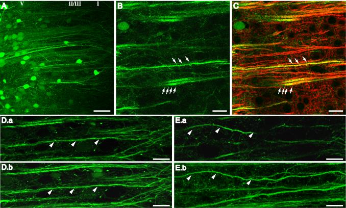
Fig. 4.
Confocal imaging of mPFC neurons transduced with AAV-GFP. (A) Clusters of fluorescent neurons surrounding the injection site in Sham-SKF mPFC. Prominent first order dendrites were long and straight, branching into tufts at the interface of layers II/III and I. (B) Representative high power image of GFP-positive dendrites in mPFC of a Les-SKF rat sacrificed at day 7 after the priming regimen. Arrows indicate the presence of dendritic spines. (C) Overlay of the image in B with the corresponding image of MAP2 immunofluorescence showing colocalization in larger dendritic shafts but not in spines (arrows). (D) Image of a single 1 μm optical section (D.a), and the corresponding reconstructed projection of 10 optical sections (D.b), of Sham-SKF mPFC. Most dendrites are visible along their entire length in both single slice images and projections (indicated by arrowheads). (E) Single 1 μm optical section (E.a), and corresponding stacked reconstruction (E.b), of dendrites in Les-SKF mPFC. Several dendrites disappeared out of the single section images (arrowheads in E.a), and could only be followed for their entire length by stacking multiple images as shown in E.b. Scale bar for A, 50 μm. Scale bar for B-E, 20 μm. Note: a magenta-green version of this figure can be viewed online as Supplementary Figure 2.

