Fig. 5.
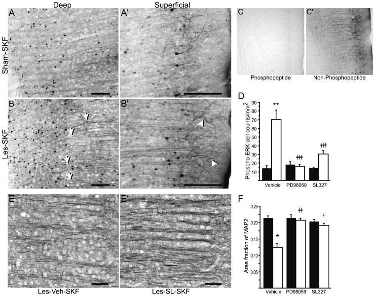
Sustained ERK phosphorylation may contribute to mPFC morphological abnormalities in D1-primed rats. Antigen retrieval was used to intensify phospho-ERK staining in Sham-SKF (A and A’) and Les-SKF (B and B’) mPFC. Note the wavy phospho-ERK-positive dendritic shafts (arrows) and enhanced immunostaining of dendrite apical tufts (arrowheads) in sections from Les-SKF (D1-primed) rat mPFC compared to that of Sham-SKF. Scale bars, 200 μm. Specificity of the antibody under antigen retrieval conditions is shown by abolition of staining using a phosphorylated (C), but not a non-phosphorylated (C’) blocking peptide. (D) MEK inhibitors administered prior to each weekly dose of SKF-38393 prevented the sustained increase in phospho-ERK observed at day 7 in D1-primed rats. Sham-lesioned (filled bars) and neonate-lesioned (open bars) animals were administered icv infusions of vehicle, PD98059 or SL327, 30 min before each weekly systemic injection of SKF-38393. Cell counts were obtained from mPFC sections immunostained for phospho-ERK without antigen retrieval and represent total counts across all cortical layers. Represented are means ± S.E.M. ** p < 0.0001 vs. sham-lesioned rats preinfused with vehicle prior to SKF-38393 (Sham-Veh-SKF); ‡‡‡ p < 0.0001 vs. D1-primed rats preinfused with vehicle (Les-Veh-SKF). (E and E’) Representative photomicrographs of MAP2 immunohistochemistry in Les-Veh-SKF and Les-SL-SKF mPFC, respectively. Scale bars, 50 μm. (F) Quantitative analysis of MAP2 immunostaining using linear area fraction measurements. The format of the graph is the same as in D. Preinfusions of either PD98059 or SL327 had no effect in sham rats treated with SKF-38393 (filled bars), but prevented the loss of linear dendritic immunostaining in lesioned rats that were primed with the agonist (open bars). * p < 0.001 vs. Sham-Veh-SKF; ‡‡ p < 0.001, and ‡ p < 0.05 vs. Les-Veh-SKF; ANOVA with Fisher’s PLSD.

