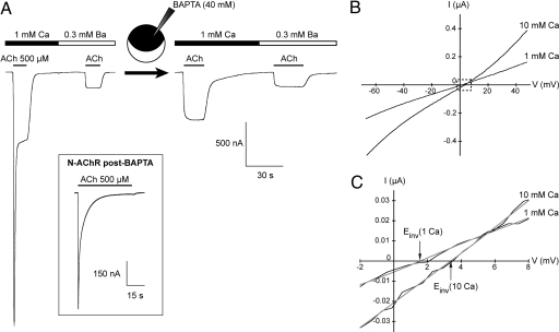
Fig. 2.
The L-AChR is permeable to calcium and shows no macroscopic desensitization. (A) All traces are from a single oocyte expressing L-AChRs. In 1 mM external CaCl2, the response to ACh displays a preeminent inward peak current. Chelating intracellular calcium by injection of BAPTA or replacing extracellular calcium by a low amount of barium (0.3 mM) eliminates this peak and results in stable responses upon continuous application of ACh. (Inset) N-AChRs display profound macroscopic desensitization upon continuous application of ACh (500 μM) even after BAPTA injection (n = 6). (B) Comparison of the I/V relationships of L-AChR responses elicited by 100 μM ACh in the presence of 1 mM or 10 mM extracellular CaCl2. Note that the L-AChR is potentiated by 10 mM extracellular calcium and that this potentiation occurs over the whole voltage range (n = 5, BAPTA-loaded oocytes). (C) Magnified view of the dash-boxed region in B showing the rightward shift of the reversal potential induced by switching from 1 mM to 10 mM external Ca2+ (1.6 mV to 3.4 mV for this cell).

