Fig. 1.
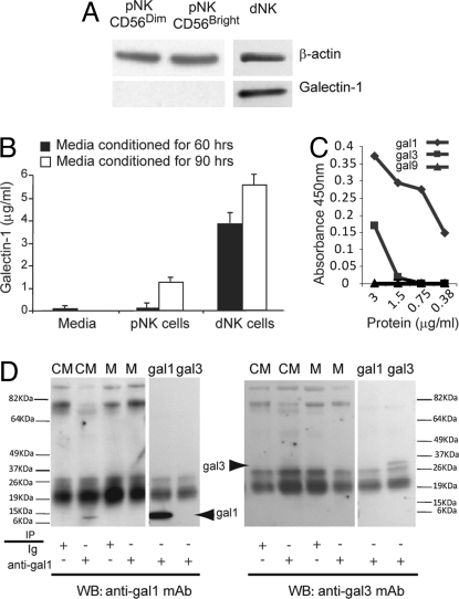
Gal1 is expressed and secreted by dNKs but not by pNKs. (A) Western blot of fresh CD56dim pNK, CD56bright pNK, and dNK lysates developed with anti-gal1 mAb (Lower) and anti-β-actin mAb (Upper). Gal1 was visualized as a 14-kDa band. Five dNK and and 5 pNK cell samples from different donors were analyzed. Similar results were obtained with decidual macrophages (data not shown). (B) Concentration of gal1 in media conditioned by CD56+CD16−CD3− dNKs and CD56+CD3− pNKs for 60 h (filled bars) and 90 h (open bars). Incubations were done in the presence of 12 ng/mL IL-15. The average of 3 independent experiments is shown. (C) Crossreactivity of the antibody used in B with recombinant human gal1 (diamonds), gal3 (squares), and gal9 (triangles) in ELISA assays. (D) Immunoprecipitation of gal1 from media conditioned by dNKs (CM), control media not exposed to dNKs (M), recombinant human gal1 (gal1), and recombinant human gal3 (gal3) with the same antibody as used in B (anti-gal1) or with an isotype control (Ig). Immunoprecipitates were resolved by Western blotting with anti-gal1 (Left) or anti-gal3 (Right) mAbs. The additional bands are derived from immunoglobulins and protein G. One experiment of 3 is shown.

