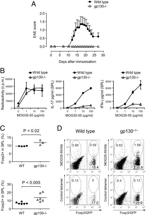
Fig. 1.
Unresponsiveness of T cells to IL-6 confers resistance to EAE caused by lack of Th17 cells and an increased T-reg response. (A) Wild-type or T cell-conditional gp130−/− mice on the Foxp3gfp.KI background were immunized with MOG35-55/CFA plus pertussis toxin and followed for signs of EAE (mean clinical score ± SEM, n = 10). (B) On day 10 after immunization, splenocytes were isolated and restimulated with MOG35-55 in vitro. The proliferative response was measured by [3H]thymidine incorporation, and the cytokine production in 48-h culture supernatants was determined by ELISA. Mean of triplicate cultures is shown. (C) Frequency of Foxp3+ T-regs in the splenic CD4+ T cell compartments of MOG35-55/CFA-immunized wild-type and gp130−/− mice as determined by the expression of Foxp3/GFP ex vivo (Upper) and after in vitro restimulation with MOG35-55 (Lower). (D) MOG35-55/IAb tetramer staining in MOG35-55-stimulated splenocytes from in vivo-sensitized wild-type and gp130−/− mice. The splenocytes were isolated on day 11 after immunization with MOG35-55/CFA followed by restimulation in vitro for 4 days. The gate was set on blasting CD4+ T cells. Representative cytograms are shown.

