Fig. 2.
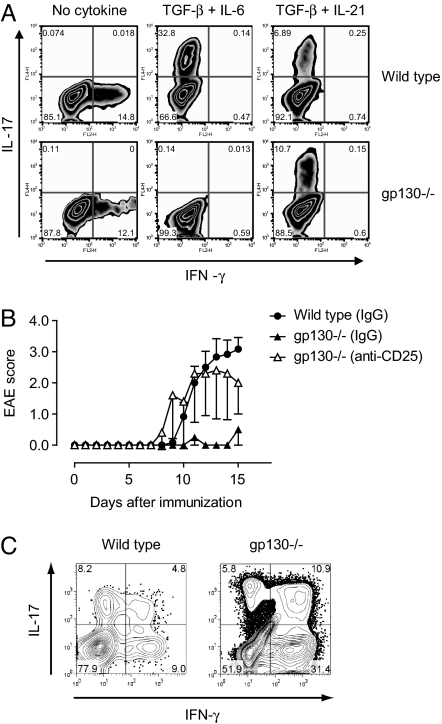
The combination of TGF-β plus IL-21 induces Th17 cells in gp130−/− mice. (A) Naïve T cells were purified from wild-type or gp130−/− mice and differentiated in vitro with either TGF-β plus IL-6 or TGF-β plus IL-21. The frequency of IL-17- and IFN-γ-positive T cells was determined by intracellular cytokine staining. (B) gp130−/− mice were either treated with control IgG (n = 3) or depleted of T-regs by treatment with a monoclonal antibody to CD25 (PC61, 2 × 0.5 mg) (8) 5 and 3 days before immunization with MOG/CFA (n = 5). As further control group, T-reg-competent wild-type mice were included (n = 6). The mean EAE score of each group is shown. Data represent 1 of 3 independent experiments. (C) At the peak of disease, mononuclear cells were isolated from the CNS of wild-type animals and T-reg-depleted gp130−/− mice followed by stimulation with PMA/ionomycin and intracellular cytokine staining for IL-17 and IFN-γ. The numbers in the quadrants of the cytograms indicate percentages of cytokine-positive cells in the CNS-derived CD4+ T cell compartment. One representative experiment is shown. Because gp130−/− mice that were not depleted of T-regs did not develop EAE, the T cellular infiltrate into the CNS of these mice was insufficient to perform intracellular cytokine staining.

