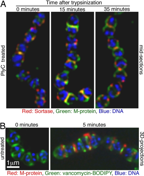
Fig. 4.
Sortase foci preferentially localize to sites of active M protein anchoring. (A) D471 cells were grown to OD600 0.5 in media containing 0.05% trypsin and either fixed immediately or washed and incubated in media without trypsin for 15 or 35 min before fixation. The cells were permeabilized, stained for sortase (red), and subsequently labeled for M protein using 3B8-FITC conjugated (green) and for DNA using DAPI (blue). (B) D471 cells were trypsinized as in A and allowed to regenerate M protein for 5 min, but were not permeabilized. The cells were stained for M protein (red) using the 10B6 monoclonal and Alexa Fluor 647 conjugate, and for DNA (blue). Vancomycin-BODIPY (green) was used to detect lipid II export regions. Images are presented as two-dimensional projection of the 3D data.

