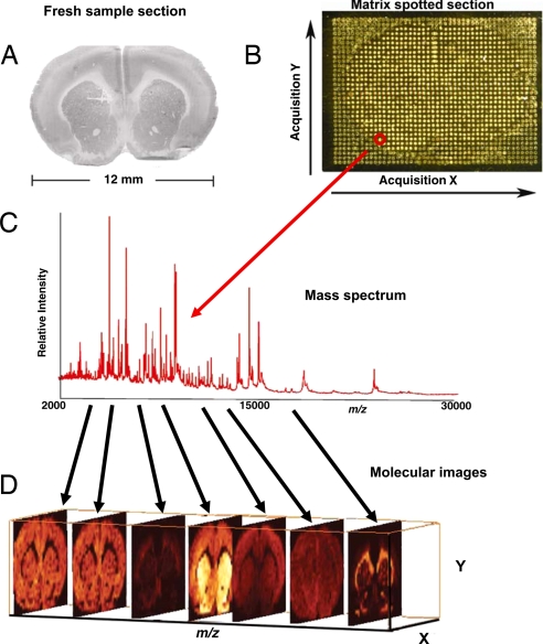
Fig. 1.
Work flow of an IMS experiment. (A) A tissue section is collected on a conductive MALDI plate. (B) Matrix is deposited in a uniform manner over the surface of the tissue. (C) Spectra are acquired from each location (pixel) over the surface of the tissue. (D) 2D ion density images are reconstructed from the spectra. Hundreds of protein images can be created from a single 12-μm-thick section of tissue from a single acquisition.

