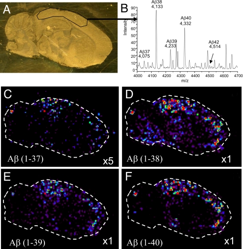
Fig. 3.
IMS allows for distinction of different molecular species of β-amyloid plaques in an Alzheimer's disease model. (A) Optical image of the mouse brain tissue section on a gold-coated MALDI target. Enclosed area is a region of high concentration of β-amyloid plaques. (B) Average spectrum from the selected region of the tissue from A. (C–F) Ion density images of four different truncations of the β-amyloid protein.

