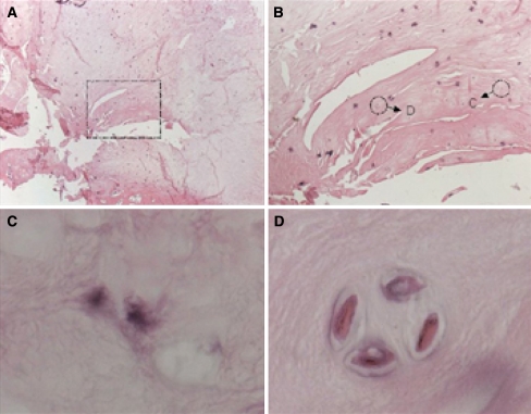
Fig. 2.
Staining of paraffin sections of the regenerated intervertebral disc 6 months following cell transplantation. BrdU containing chondrocytes were detected and stained by immunohistochemical procedures using DAB as the chromogen. Sections were counterstained by Eosin. BrdU positive cells are colored black. a Nucleus regenerate overview (25×), b BrdU stained transplanted cells (200×), c, d single BrdU stained transplanted chondrocytes, pericellular de novo synthesis of nucleus matrix (1,000×)

