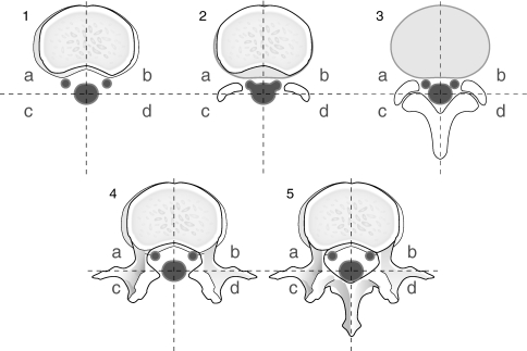
Fig. 2.
The amount and location of peridural scar was evaluated according to the grading system described by Ross et al. by using a score of 0–4. Five axial MRI slices/patient were available for evaluation (1–5). Two slices above the disc (1, 2), one in the disc level (3) and two below the disc (4, 5). Each slice was divided into four quadrants (a–d)

