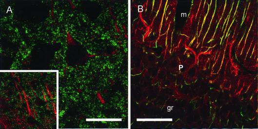
Figure 3.
Immunofluorescence labeling of cerebral cortex for Nav1.6 and synaptic vesicles and of cerebellar cortex for Nav1.6 and glial cells. Single confocal sections. (A) Cerebral cortex labeled for Nav1.6 (red) and SV2 (green). Apical dendrites of neurons were labeled for Nav1.6 within the dendrite. SV2 labeling was throughout the neuropil and outlined the cell bodies and large dendrites of the neurons. (Inset) Deconvolved image of cortical neurons (lower magnification than in A). (B) Cerebellar cortex labeled for Nav1.6 (red) and GFAP (green). Nav1.6 labeling was intense in apical dendrites of Purkinje cells in the vertically oriented GFAP-positive radial glial cell processes (yellow) and was diffusely present in the molecular layer (m). Glial cell processes that were GFAP-positive in the granule cell layer (gr) were not labeled for Nav1.6. Purkinje cell bodies (P layer) were lightly labeled in the cytoplasm. (Bars = 20 μm for A, 75 μm for Inset, and 50 μm for B.)

