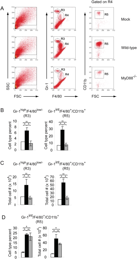
Figure 5. Recruitment of inflammatory monocytes/macrophages to the SARS-CoV infected lung is delayed in MyD88−/− mice as compared to WT mice.
10 wk old B6 WT or MyD88−/− mice were inoculated intranasally with PBS or 105 pfu rMA15. (A) At 2 dpi, lung leukocytes were isolated as described in Materials and Methods and analyzed by flow cytometry. Histograms are representative of three mice. Two independent experiments gave similar results. (B) Percent inflammatory monocytes/macrophages of total lung leukocytes isolated from mock (□), rMA15-infected WT (▪), or rMA15-infected MyD88−/− (▪) mice at 2 dpi. (C) Total numbers of inflammatory monocytes/macrophages isolated from mock (□), rMA15-infected WT (▪), or rMA15-infected MyD88−/− (▪) mice at 2 dpi. (D) Percent inflammatory monocytes/macrophages of total lung leukocytes (left panel) and total numbers of inflammatory monocytes/macrophages (right panel) isolated from mock (□), rMA15-infected WT (▪), or rMA15-infected MyD88-/-n (▪) mice at 4 dpi.

