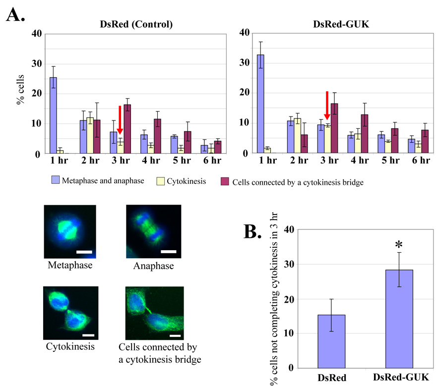
Fig. 3. Delayed cytokinesis in cells over-expressing the DsRed-GUK.
(A) HeLa cells expressing control DsRed or DsRed-GUK were arrested in the M phase by double thymidine block and nocodazole treatment. At the indicated hours after the washout of nocodazole, cells were fixed and stained for tubulin and DNA. Mitotic cells were counted at the metaphase, anaphase, cytokinesis, and interphase when they were still connected by a “cytokinesis bridge”. The data reflect mean ± SD from three independent experiments. The representative images are shown in the bottom panels. Bar 10 µm. (B) Percentage of cells not completing cytokinesis at three hours after nocodazole washout. The value is the percentage of cytokinesis at three hours divided by the percentage of metaphase and anaphase cells at one hour. Data are mean ± SD from three independent experiments. *P < 0.05.

