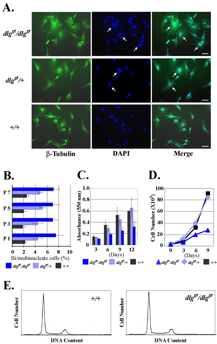
Fig. 4. Multinuclearity and reduced proliferation of dlg mutant MEFs.
(A) Multinucleated cells in dlggt/dlggt MEFs. Homozygous dlggt/dlggt, heterozygous dlggt/+, and wild type +/+ MEFs obtained from littermates were plated on glass coverslips, and stained for β-tubulin (green) and DNA with DAPI (blue). Multinucleated cells are indicated by arrows. Bar 50 µm. (B) Quantification of multinucleated cells. At the indicated number of passages, the MEFs were analyzed for the number of multinucleated cells. Three independent preparations of MEFs for homozygous, heterozygous, and wild type were used for the analysis. Data are mean ± SD. At least 225 cells for each sample were counted. (C) Proliferation assay of MEFs by crystal violet metod. Passage 1 MEFs were plated in the 96 well plates and the cell number was quantified by the crystal violet method at the indicated days after plating. Three independent preparations of MEFs for homozygous, three for heterozygous, and two for the wild type were derived from littermate embryos and the mean ± SD values are shown. (D) Proliferation of MEFs by direct cell counting. 3×10⁵ MEFs were passaged every 3 days in a 6 well plate. The cell numbers were counted at each passage point. (E) Flow cytometry of the DNA content of the wild type and dlggt/dlggt MEFs.

