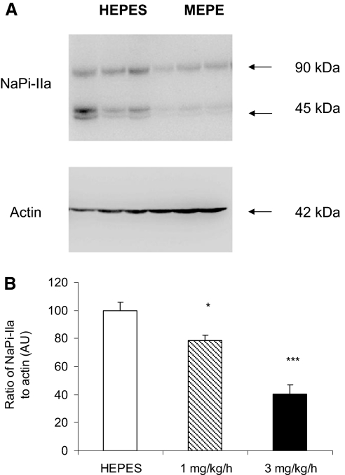
Figure 4.
Western blot analysis of NaPi-IIa protein in the kidneys of rats infused with HEPES or MEPE. (A) Representative Western blot of NaPi-IIa in BBM vesicles prepared from HEPES and 3 mg/kg per h MEPE-treated rats. (B) Quantification of NaPi-IIa protein relative to β-actin. Data are means ± SEM of Western blots performed on six individual BBM vesicle samples from each experimental group. The abundance of NaPi-IIa is given as a ratio of NaPi-IIa protein to β-actin protein in arbitrary units (AU). *P < 0.05 and ***P < 0.001 compared with HEPES control using an ANOVA with post hoc comparisons performed using the Bonferroni multiple comparisons test.

