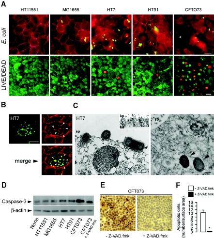
Figure 2.
Effects of UPEC on the integrity of confluent renal mpkIMCD cell layers. (A) E. coli isolates (5 × 10⁵ bacteria/well) were added (3 h) to the apical side of confluent mpkIMCD cells grown on 1-μm-pore-size permeable filters, then fixed and processed for indirect immunofluorescence using an anti–ZO-1 antibody (in purple), and then stained with phalloidin (in red) to visualize F-actin. Cells were also stained with LIVE/DEAD fluorescent dye staining (red, dead cells; green, viable cells). CFT073 but not HT7 and HT91 altered the integrity of the cell layer with numerous dead cells (*). Bars = 10 μm. (B) Indirect immunofluorescence of cells grown on glass slide showing actin rearrangements (in red) forming close contacts (arrowhead) with an adherent HT7 bacteria (in green, and merge image in yellow). (C) Transmission electron micrographs of adherent HT7 bacteria to the apical membrane of mpkIMCD cells (left) and internalized bacteria (arrowheads; right). Bars = 1 μm. (D) Western blot analysis of active caspase-3 expression in nonpathogenic and uropathogenic E. coli strains. (E and F) Illustrations (E) and number of caspase-3–positive stained cells (F) incubated with CFT073 and without or with the pan-caspase inhibitor Z-VAD.fmk (20 μM, 30 min). Bars are means ± SEM; n = 3 independent experiments. *P < 0.05 between groups. Bar = 10 μm.

