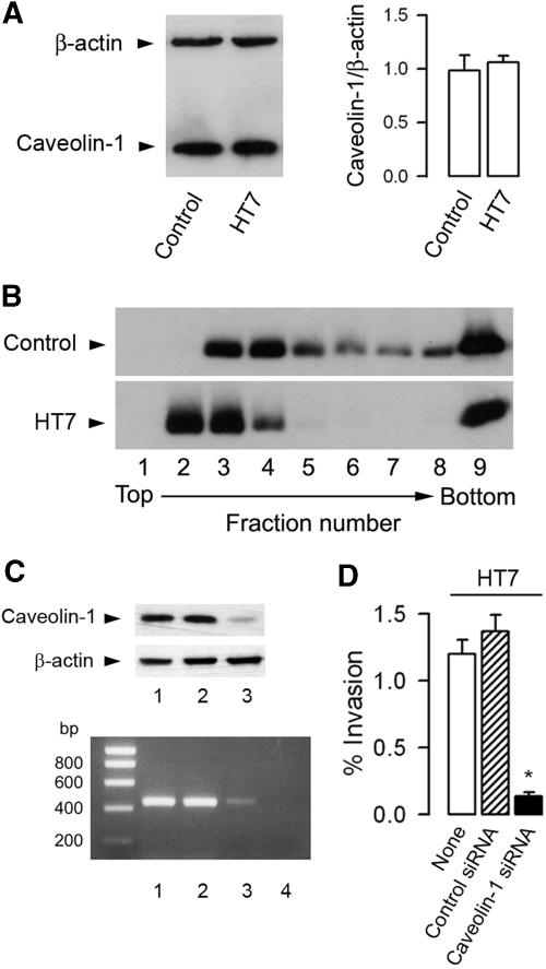
Figure 4.
Caveolin-1 requirement for internalization of UPEC in mpkIMCD cells. (A) Western blot analysis of caveolin-1 and β-actin in mpkIMCD cells incubated without (control) or with HT7 (5 ± 105 bacteria/well, 3 h). Bars are the mean ratio values ± SEM of densitometric analyses of caveolin-1 over β-actin–labeled bands from three to four separate cultures. (B) Western blot analysis of caveolin-1 in DRM fractions fractionated by flotation centrifugation gradient in uninfected and HT7-infected mpkIMCD cells. (C) Caveolin-1 mRNA (bottom) and protein (top) expressions in uninfected mpkIMCD cells (lane 1) or cells transfected with a negative control siRNA (lane 2), a caveolin-1 siRNA (lane 3), or with non–reverse-transcribed caveolin-1 siRNA (lane 4). (D) Nontransfected cells (None) and transfected cells with caveolin-1 siRNA or negative control siRNA (control siRNA) were incubated with HT7 (5 ± 105 bacteria/well, 3 h) before invasion assay. Bars (means ± SEM) represent the percentage of internalized bacteria. *P < 0.05 versus None values.

