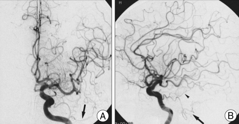
Fig. 2.
Patient 3. Left internal carotid angiograms, anteroposterior view (A) and lateral view (B), showing the persistent trigeminal artery variant (arrow) anastomosing the cavernous portion of the internal carotid artery to the superior cerebellar artery and anterior inferior cerebellar artery (arrow head).

