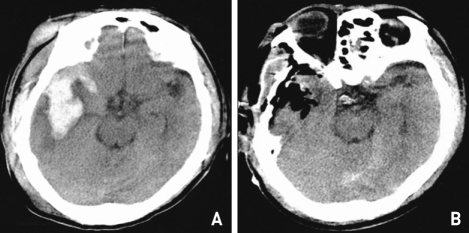
Fig. 2.
A : Preoperative brain computed tomography (CT) scan showing a large scalp hematoma and fatal intracerebral hematoma in the right temporoparietal region, centered on the sylvian fissure, contacting the dura mater and extending to the ventricles. B : Postoperative CT scan showing evidence of right frontotemporal cranioplasty and hematoma removal.

