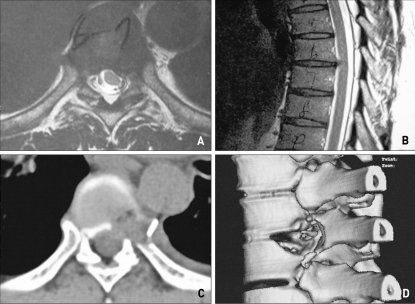
Fig. 3.
A case of 47 years old male patient with thoracic herniated nucleus pulposus. Preoperative MRI shows extruded disc compressing nerve root at the level of T6-7, left side (A, B). T2-weighted axial (A) and T2-weighted sagittal (B). Postoperative CT scan demonstrates satisfactory decompression of the T7 vertebra and rib head with disc space at T6-7 level (C, D).

