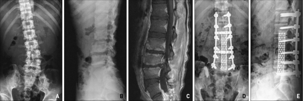
Fig. 2.
Plain radiographs of pyogenic spondylitis with scoliosis, bony erosion at L3,4,5 (A, B). Magnetic resonance imaging of pyogenic spondylitis with severe enhanced vertebral body involvement, L3/4/5 (C). Postoperative plain radiograph of the combined approach (D, E). Mesh insertion and screw fixation were performed. The patient presented with lower extremity weakness. After four months of treatment, the patient could walk without assistance.

